When your doctor recommends imaging to check your internal organs, you might hear the terms sonography and ultrasound used interchangeably. But are they really the same thing? Or is there a subtle distinction?
If you’ve ever wondered about sonography vs ultrasound, you’re not alone. While closely related, these terms describe different aspects of the same diagnostic technique. In this article, we’ll explain the difference between ultrasound and sonography, how each term is used, and what to expect during a diagnostic ultrasound.
What Is Ultrasound?
Ultrasound refers to the technology that uses high-frequency sound waves to create images of structures inside your body. The sound waves are emitted by a handheld probe, bounce off tissues and organs, and are then captured to produce real-time images on a monitor.
Ultrasound is widely used in medicine because it is:
- Non-invasive
- Painless
- Free from ionizing radiation (unlike X-rays or CT scans)
- Quick and highly effective for soft tissue imaging
Common uses of ultrasound imaging explained include:
- Monitoring pregnancy and fetal development
- Checking abdominal organs (liver, kidneys, gallbladder, pancreas)
- Evaluating heart function (echocardiography)
- Examining blood flow in arteries and veins
- Guiding biopsies and minimally invasive procedures
What Is Sonography?
Sonography refers to the process of performing an ultrasound scan — that is, the act of using the ultrasound machine to acquire and interpret images. In simple terms, sonography is the practice, while ultrasound is the technology.
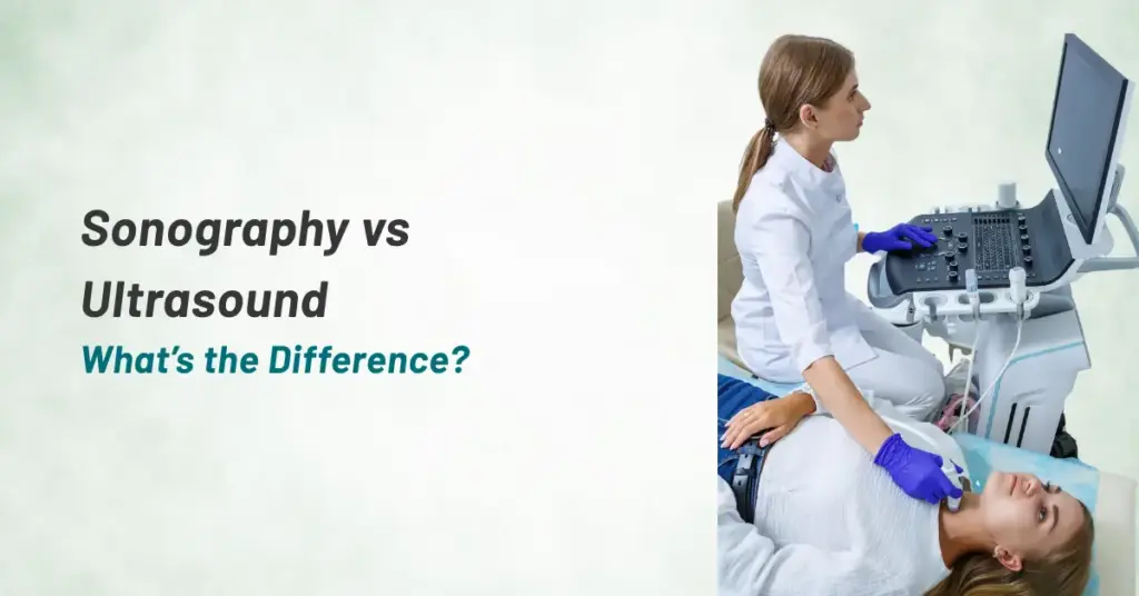
A sonographer is a trained healthcare professional who performs the scan, positions the probe, adjusts settings for optimal imaging, and often provides preliminary observations.
So when people ask what is sonography test, it’s helpful to think of it as the application of ultrasound technology to examine a specific part of the body for diagnostic purposes.
Key Differences Between Sonography and Ultrasound (Quick Table)
When people search for sonography vs ultrasound, the confusion is understandable. Both terms are closely connected, but they are not exactly the same.
| Aspect | Ultrasound | Sonography |
| Meaning | The technology using sound waves | The process of performing the scan |
| Focus | Equipment and imaging method | Practical application |
| Performed by | Doctors or technicians | Trained sonographer |
| Purpose | Produces internal images | Captures and interprets images |
In simple terms, ultrasound is the tool, while sonography is the method used to carry out the test. This distinction helps answer the common question about the difference between ultrasound and sonography, especially in clinical settings where accuracy matters.
When Do Doctors Recommend Ultrasound?
Doctors recommend ultrasound as a first-line diagnostic tool because it is safe, quick, and provides real-time images. If you’re wondering what is sonography test, it refers to the actual procedure where a trained professional uses ultrasound technology to examine specific parts of the body.
You may be advised to undergo this test in situations such as:
- Abdominal discomfort to evaluate organs like the liver, kidneys, or gallbladder
- Pregnancy monitoring to track fetal growth and development
- Swelling, lumps, or pain to identify underlying causes
- Heart function assessment, where ultrasound helps visualize heart movement
- Blood flow concerns, especially in arteries and veins, to detect clots or narrowing
Because sonography is non-invasive and does not involve radiation, it is often the preferred choice for both diagnosis and follow-up.
Is Ultrasound Safe?
One of the most common concerns patients have is about safety. The good news is that ultrasound is considered extremely safe when performed correctly. It uses sound waves, not radiation, which makes it suitable even for repeated use.
Whether you are comparing sonography vs ultrasound or preparing for your first scan, it helps to know that:
- The procedure is non-invasive and painless
- There are no known harmful side effects
- It is widely used during pregnancy without risk to the baby
- Trained professionals ensure proper technique and accurate results
Understanding the difference between ultrasound and sonography can also reassure patients that both refer to a reliable and well-established diagnostic approach. The technology and the process work together to provide clear insights into your health.
Most people complete the test comfortably within a short time and can return to normal activities immediately. If you still have concerns, your doctor or sonographer can guide you through the process step by step, helping you feel more confident and informed.
Sonography vs Ultrasound: Are They the Same?
Here’s the key point: ultrasound refers to the sound waves and the technology itself, while sonography refers to the procedure of using ultrasound for medical imaging. This clearly explains the ultrasound and sonography difference in a clinical context.
Think of it this way:
- Ultrasound = the tool/technology
- Sonography = the use of the tool in practice
They’re often used interchangeably in casual conversation because they’re so closely related. However, in medical terminology, there is a distinction between the two.
Types of Ultrasound Scans
To better understand sonography vs ultrasound, it helps to know that sonography is performed using different types of ultrasound scans, depending on the part of the body being examined. Some common examples include:
Abdominal Ultrasound
Used to evaluate the liver, gallbladder, spleen, kidneys, pancreas, and bladder.
Obstetric and Gynecologic Ultrasound
Used to monitor pregnancy, check the uterus and ovaries, and assess fetal health.
Echocardiography
An ultrasound of the heart to assess structure, function, and blood flow.
Vascular Ultrasound
Evaluates blood flow in arteries and veins to detect blockages or clots.
Musculoskeletal Ultrasound
Examines muscles, tendons, and joints for injuries or inflammation.
Each of these scans involves diagnostic ultrasound technology and is performed through the practice of sonography.
Why Are These Terms Confused?
Part of the confusion between sonography vs ultrasound comes from the fact that the terms are often used synonymously in healthcare settings. For example:
- A doctor may say, “Schedule an ultrasound,” referring to the test.
- The technician performing it may say they’re “doing a sonography.”
Both refer to the same procedure, just from slightly different perspectives.
What Happens During a Sonography/Ultrasound?
If your doctor recommends a diagnostic ultrasound, here’s what to expect:
- You may be asked to change into a gown.
- A water-based gel is applied to the area being scanned.
- The sonographer moves a handheld transducer over your skin, sending sound waves into your body.
- Images appear on a monitor in real time, and the sonographer may capture specific views for analysis.
- The test is painless and typically takes 15–45 minutes.
After the procedure, a radiologist reviews the images and sends a report to your doctor, who will discuss the findings with you.
Advantages of Ultrasound/Sonography
Some of the biggest benefits of ultrasound and sonography include:
- No radiation exposure
- Widely available and cost-effective
- Real-time imaging
- Non-invasive with no known risks
This makes it a preferred imaging method for many diagnostic and monitoring purposes.
Final Thoughts
So, what’s the difference between sonography and ultrasound? Simply put:
- Ultrasound is the technology — the sound waves and the machine.
- Sonography is the process — using the technology to perform the scan.
Both terms are closely related and often used interchangeably, but understanding the distinction can help you communicate more clearly with your healthcare provider.
Whether you’re undergoing an abdominal scan, pregnancy check, or vascular assessment, rest assured that both ultrasound technology and the skilled practice of sonography play vital roles in providing you with accurate, safe, and timely diagnostic information.
If you have concerns or questions about your upcoming scan, don’t hesitate to ask your doctor or sonographer. Knowing what to expect helps you feel more comfortable and informed about your healthcare choices.