Prashanth Institute of Neurology & Neurosciences offers super-specialty care for all kinds of neurological disorders. We have a dedicated team of neurologists and neurosurgeons offering a wide range of treatments for neurological disorders. Our neuro team works in an integrated approach along with intensivists and rehabilitation specialists to provide the best possible treatment. The surgical team provides cutting edge technology and treatment for the entire spectrum of neurological diseases. We offer 24 x 7 services for trauma and strokes with a separate ICU. Our team of experts offers the best neuroscience care, such as inpatient and outpatient treatment, neuro-diagnostic, interventional procedures, and surgery.
We have a complete spectrum of treatment capabilities in neurosciences and can truly select what modality would be best for the patient. Prashanth Hospitals has dedicated units, which are further streamlined into various sub-disciplines of Neuroscience. We have the best brain specialists in Chennai.
• Expert care for all neurological disorders
• Headache clinic
• Treatment for epilepsy, movement disorders, and demyelinating disorders
• 24 x 7 World-class Stroke Clinic
• EEG and nerve conduction facilities
• Vertigo/Parkinson’s/Dementia Clinic
• Neurosurgery
• Advanced brain and spine care center
• Minimally invasive neurosurgical procedures of the brain and spine
• Epilepsy surgery
• Skull-based surgery
• Cerebrovascular surgery
• Deep Brain Stimulation
• Dedicated physiotherapy
Prashanth Hospitals is one of the best neuro hospitals in Chennai. We take every measure to ensure that patients and their families are comfortable, with our experts using a team approach to deliver the most advanced care possible with the best neuro specialists in Chennai.
Our expert Neurology team deals with conditions such as dementia, epilepsy, movement disorders, chronic pains, sleep disorders, headaches, multiple sclerosis, and numerous other neuromuscular diseases. At Prashanth, we have the best neurologists in Chennai treating patients with premier care.
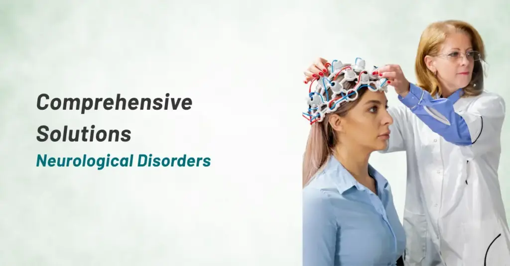
An electroencephalogram (EEG) is a noninvasive test that records electrical patterns in the brain. EEG measures voltage fluctuations resulting from ionic current flows within the neurons of the brain. An EEG measures electricity that your brain makes; it does not measure thoughts or feelings, and it does not send any electricity into your brain. In a clinical context, EEG refers to the recording of the brain’s spontaneous electrical activity over a short period of time, usually 20–40 minutes, as recorded from multiple electrodes placed on the scalp. In neurology, the main diagnostic application of EEG is in the case of epilepsy, as epileptic activity can create clear abnormalities on a standard EEG study. Secondary clinical use of EEG is in the diagnosis of coma, encephalopathies, and brain death. EEG is also used for studies of sleep and sleep disorders where recordings are typically done for one full night, sometimes more.
Nerve conduction study (NCS) is a test commonly used to evaluate the function, especially the ability of electrical conduction, of the motor and sensory nerves of the human body. Nerve conduction velocity (NCV) is a common measurement made during this test. NCS measures how fast and how strong the electrical activity is in a nerve. The test can tell whether a nerve has been damaged.
Nerve conduction studies are used mainly for evaluation of paresthesias (numbness, tingling, burning) and/or weakness of the arms and legs. The type of study required is dependent in part by the symptoms presented. A physical exam and a thorough history also help to direct the investigation.
Some common disorders that can be diagnosed by nerve conduction studies:
• Peripheral neuropathy
• Carpal tunnel syndrome
• Ulnar neuropathy
• Guillain-Barré syndrome
• Facioscapulohumeral muscular dystrophy
• Spinal disc herniation
• Cubital Tunnel Syndrome
• Tarsal Tunnel Syndrome
• Vertigo/ Parkinson’s/ Dementia Clinic
NCS can also help your doctor see how well you’re recovering from a nerve injury.
Electromyography (EMG) is a technique for evaluating and recording the electrical activity produced by skeletal muscles. It is performed using an instrument called an electromyograph, to produce a record called an electromyogram. It detects the electrical potential generated by muscle cells when these cells are electrically or neurologically activated. The signals can be analyzed to detect medical abnormalities, activation level, and recruitment order or to analyze the biomechanics of human or animal movement.
The Headache Clinic at Prashanth Hospitals treats patients with the understanding that headache is not only a biological or neurological disorder but a disorder influenced by psychological, social, and environmental stressors. Headache disorders, particularly chronic ones such as a chronic migraine can be highly debilitating, impacting domestic and professional responsibilities and emotional functioning. Keeping this in mind, we implement the most appropriate approach towards treating a headache.
A headache or cephalalgia is pain anywhere in the region of the head or neck. It can be a symptom of a number of different conditions. The brain tissue itself is not sensitive to pain because it lacks pain receptors. Rather, the pain is caused by disturbance of the pain-sensitive structures around the brain. Nine areas of the head and neck have these pain-sensitive structures, which are the cranium (the periosteum of the skull), muscles, nerves, arteries and veins, subcutaneous tissues, eyes, ears, sinuses, and mucous membranes. There are a number of different classification systems for headaches. Headache is a non-specific symptom, which means that it has many possible causes.
Treatment for headache depends on the underlying etiology or cause, but commonly involves analgesics.
These factors are thought to contribute to tension headaches:
• Stress
• Depression
• Anxiety
• Bad posture
• Staying in one position for a long time
• Working in an awkward position for a long time
• Clenching one’s jaw
Epilepsy is a diverse set of chronic neurological disorders characterized by seizures. Some definitions of epilepsy require that seizures are recurrent and unprovoked but others require only a single seizure combined with brain alterations which increase the chance of future seizures. In many cases, a cause cannot be identified. However, factors that are associated include brain trauma, strokes, brain cancer, and drug and alcohol misuse among others. Epileptic seizures result from abnormal, excessive, or hypersynchronous neuronal activity in the brain.
Epilepsy becomes more common as people age. The onset of new cases occurs most frequently in infants and the elderly. As a consequence of brain surgery, epileptic seizures may occur in recovering patients. Epilepsy is usually controlled, but not cured, with medication. More than 30% of people with epilepsy do not have seizure control even with the best available medications. Surgery may be considered in difficult cases. Not all epilepsy syndromes are life long – some forms are confined to particular stages of childhood. Epilepsy should not be understood as a single disorder, but rather as syndromic with vastly divergent symptoms, all involving episodic abnormal electrical activity in the brain and numerous seizures.
DBS is a surgical procedure where slender electrical wires are accurately placed in deep locations within the brain and connected to a battery that is placed under the skin of the upper chest. Using these, the doctors are able to modulate the intricate neuronal circuitry in the brain. The amount of current that is sent can be controlled with a remote controller. Following DBS, the patient experiences a significant reduction in the movements, the fluctuations are smoothened out, and the dose of medicines required can be reduced. Simply put, the patient can expect a good improvement in the quality of life!
• It has proven very useful in conditions like generalized dystonia where there is a painful spasmodic muscle movement occurring in many different muscles all at the same time
• Extremely effective in essential tremor
• Proven to be effective in patients with epilepsy
• Surgical therapies may be considered during the course of Parkinson’s disease especially if symptoms cannot be adequately controlled with medication.
Movement disorders are neurological disorders in which there is either an excess of movement or a paucity of movement that is not due to weakness or paralysis. Movement disorders are associated with changes in the brain cells that help us move. These brain cell changes can cause extra unwanted involuntary or excessive movements, called hyperkinesia. Other changes in brain cell function can cause a lack of automatic and purposeful movements, not related to weakness or spasticity, called hypokinesia. A complete medical history and careful thorough neurological examination are required to make an accurate diagnosis. Movement disorders, once correctly diagnosed, usually respond to treatment.
It is a disease of the nervous system in which the myelin sheath of neurons is damaged. This damage impairs the conduction of signals in the affected nerves. In turn, the reduction in conduction ability causes a deficiency in sensation, movement, cognition, or other functions depending on which nerves are involved. Some demyelinating diseases are caused by genetics, some by infectious agents, some by autoimmune reactions, and some by unknown factors. Organophosphates, a class of chemicals which are the active ingredients in commercial insecticides such as sheep dip, weed-killers, and flea treatment preparations for pets, etc., will also demyelinate nerves. Neuroleptics can also cause demyelination. The precise mechanism of demyelination is not clearly understood but there is good evidence that the body’s own immune system is at least partially responsible. Acquired immune system cells called T-cells are known to be present at the site of lesions. Other immune system cells called Macrophages (and possibly Mast cells as well) also contribute to the damage.
Treatment typically involves improving the patient’s quality of life. This is accomplished through the management of symptoms or slowing the rate of demyelination. Treatment can include medication, lifestyle changes (i.e. quit smoking, adjusting daily schedules to include rest periods and dietary changes), counseling, relaxation, physical exercise, patient education, and in some cases, deep brain thalamic stimulation. The progressive phase of myelin sheath appears to be driven by the innate immune system, which may directly contribute to the neurodegenerative changes that occur in the progressive myelin sheath. Until now, there are no therapies that specifically target innate immune cells in the myelin sheath. As the role of innate immunity in the myelin sheath becomes better defined, it may be possible to better treat it by targeting the innate immune system.
Our 24-hour stroke clinic is managed by expert and experienced neurologists. A stroke, or cerebrovascular accident (CVA), is the rapid loss of brain function due to disturbance in the blood supply to the brain. This can be due to ischemia (lack of blood flow) caused by blockage (thrombosis, arterial embolism), or a hemorrhage. As a result, the affected area of the brain cannot function, which might result in an inability to move one or more limbs on one side of the body, inability to understand or formulate speech, or an inability to see one side of the visual field.
A stroke is a medical emergency and can cause permanent neurological damage and death. Risk factors for stroke include old age, high blood pressure, previous stroke or transient ischemic attack (TIA), diabetes, high cholesterol, tobacco smoking, and atrial fibrillation. High blood pressure is the most important modifiable risk factor of stroke. It is the second leading cause of death worldwide.
If the patient with ischemic stroke reaches hospital within the window period of 4.5 hours, he/she can be treated with thrombolysis (also known as a “clot buster”), and there is possibility of complete recovery. Even after 4.5 hours suitable patient can undergo mechanical thrombectomy to remove the clot. Some hemorrhagic strokes benefit from neurosurgery. Treatment to recover any lost function is termed stroke rehabilitation, ideally in a stroke unit and involving health professions such as speech and language therapy, physical therapy, and occupational therapy.
• Administration of antiplatelet drugs such as aspirin and dipyridamole
• Control and reduction of high blood pressure
• Use of statins - Selected patients may benefit from carotid endarterectomy and the use of anticoagulants
• During minimally invasive neurosurgery, the diseased portion of the brain (including the fluid-filled ventricles), or spine is reached through the smallest possible incision or “keyhole’.
• With the insertion of a small endoscope equipped with a tiny camera, through a “key-hole” incision in the spine, scalp, or nostrils, minimally invasive neurosurgeons can treat a variety of neurological conditions.
• Results are faster and simpler with greatly reduced postoperative complications, minimal brain trauma, faster recovery, and less pain and scarring.
A certain group of epileptic patients who are refractory to medical treatment will be cured by epilepsy surgery after proper evaluation and work up. We provide all types of epilepsy surgery for suitable patients.
This is the most common way to remove pituitary tumors. Transsphenoidal means that the surgery is done through the sphenoid sinus, through the nostril a hollow space in the skull behind the nasal passages and below the brain. The back wall of the sinus covers the pituitary gland.
Less commonly, for larger or more complicated pituitary tumors, a craniotomy may be needed. In this approach, the surgeon operates through an opening in the front and side of the skull. The surgeon has to carefully work beneath and between the lobes of the brain to reach the tumor. It is actually safer for large and complex lesions because it provides better visualization and control of important nerves and blood vessels.
Arteriovenous malformations (AVMs) in the brain can cause seizures and hemorrhage. Many times they present as an acute emergency. In the spinal cord, they may present as back pain or progressive weakness, sometimes as acute hemorrhage and weakness. They require evaluation with a CT brain with an angiogram followed by meticulous microscopic excision.
Clipping is a surgical procedure performed to treat a balloon-like bulge of an artery wall known as an aneurysm. As an aneurysm grows, it becomes thinner and weaker. It can become so thin that it leaks or ruptures, releasing blood into the spaces around the brain – called a subarachnoid hemorrhage, a life-threatening problem. A tiny clip is placed across the neck of the aneurysm to stop or prevent an aneurysm.
Ruptured Aneurysms burst open and release blood into the space between the brain and skull, called a subarachnoid hemorrhage (SAH). Vasospasm is a common complication of SAH, which must be closely managed after treatment to prevent stroke.
Unruptured Aneurysms may not cause symptoms and are typically detected during routine testing. People with a family history of brain aneurysms should have a screening test (CT or MR angiogram). The risk of aneurysm rupture is about 1% per year of the remaining life span but may be higher or lower depending on the size and location of the aneurysm. However, when rupture occurs, the risk of death is 40%, and the risk of disability is 80%.
Neurosurgery in pediatric patients requires a lot of experience and meticulous handling of the growing brain and spinal cord. Many of the congenital abnormalities in children from newborn to adult children, like Hydrocephalus, spinal dysraphism, craniosynostoses, and Craniovertebral junction anomalies require timely identification and proper management. Apart from pediatric brain tumors like gliomas, medulloblastoma, craniopharyngioma, and pineal region, tumors should be identified earlier and treated. The earlier the treatment, the longer are the chances of survival.
In coordination with neuro physicians and stroke ICU intensivists, lifesaving procedures like Decompressive craniectomy, EVD, and evacuation of hematoma in hypertensive ICH and CVT are done. These procedures offer a timely intervention in stroke management thereby avoiding mortality, reducing the morbidity, and improving the overall quality of life. We make sure that every patient under our care receives a high standard of stroke management services.
Electroencephalography (EEG) is the recording of electrical activity along the scalp. An electroencephalogram (EEG) is a noninvasive test that records electrical patterns in the brain. EEG measures voltage fluctuations resulting from ionic current flows within the neurons of the brain. An EEG measures electricity that your brain makes; it does not measure thoughts or feelings, and it does not send any electricity into your brain. In a clinical context, EEG refers to the recording of the brain’s spontaneous electrical activity over a short period of time, usually 20–40 minutes, as recorded from multiple electrodes placed on the scalp. In neurology, the main diagnostic application of EEG is in the case of epilepsy, as epileptic activity can create clear abnormalities on a standard EEG study. Secondary clinical use of EEG is in the diagnosis of coma, encephalopathies, and brain death. EEG is also used for studies of sleep and sleep disorders where recordings are typically done for one full night, sometimes more.
A nerve conduction study (NCS) is a test commonly used to evaluate the function, especially the ability of electrical conduction, of the motor and sensory nerves in the human body. Nerve conduction velocity (NCV) is a common measurement made during this test. Nerve conduction studies are used mainly for evaluation of paresthesia (numbness, tingling, burning) and/or weakness of the arms and legs. The type of study required is dependent in part by the symptoms presented. A physical exam and a thorough history also help to direct the investigation. Some of the common disorders that can be diagnosed by nerve conduction studies are:
• Peripheral neuropathy
• Carpal tunnel syndrome
• Ulnar neuropathy
• Guillain-Barre syndrome
• Facioscapulohumeral muscular dystrophy
• Spinal disc herniation
• Cubital Tunnel Syndrome
• Tarsal Tunnel Syndrome
Electromyography (EMG) is a technique for evaluating and recording the electrical activity produced by skeletal muscles. It is performed using an instrument called an electromyograph, to produce a record called an electromyogram. It detects the electrical potential generated by muscle cells when these cells are electrically or neurologically activated. The signals can be analyzed to detect medical abnormalities, activation level, and recruitment order or to analyze the biomechanics of human or animal movement.
What is neurosurgery?
Neurosurgery is the surgical treatment of the nervous system and its coverings. The neurosurgeons operate on the brain, spinal cord, skull and scalp, and the spine (bony vertebral column). This specialty includes many different types of disorders, including epilepsy, tumors, vascular disorders (disorders of blood vessels), and spine tumors, among many others.
What is the difference between a neurologist and a neurosurgeon?
A neurologist and a neurosurgeon are similar to a cardiologist and a cardiac surgeon; the neurosurgeon employs surgical means to treat neurological disorders, while the neurologist identifies neurological disorders through diagnostic testing and uses medical (pharmacologic) means to treat them.
What is epilepsy?
Epilepsy is a brain disorder involving recurrent seizures or temporary alteration in one or more brain functions. An adult with uncontrolled epilepsy may not be able to work or drive a car. In children, epilepsy can interfere with learning and other cognitive functions. Epilepsy begins anywhere between the ages of 3 and 14 years and continues indefinitely.
What are seizures?
A seizure, or convulsion, can be a sudden, violent, uncontrollable contraction of a group of muscles caused by abnormal electrical activity in the brain. A seizure can also be more subtle, consisting of only a brief “loss of contact” (victim seems to be daydreaming) or a repetitive body movement. Epileptic seizures can be classified into two broad groups—generalized and partial. Generalized seizures affect the whole body, causing a loss of consciousness, and may arise over a wide area of the brain. Partial seizures, during which consciousness may be retained, are usually caused by damage to a more limited area of the brain. Seizures can be treated with anticonvulsant medications or surgery.
Is epilepsy the only cause of seizures?
There are a number of common causes of seizures which include:
• Injury or trauma to the head
• Infection
• Brain tumor (30 to 40 percent of patients with brain tumors have a seizure)
• High fever or heatstroke
• Diabetes (seizures can occur when the blood sugar level is too low)
There may be other causes of seizures, including unlikely diseases and medications. Furthermore, the causes may vary based on age and gender of the affected person, as well as on the specific characteristics of the symptom such as quality, time course, aggravating factors, relieving factors, and associated complaints. It is important to see a doctor for a thorough evaluation to determine a seizure’s cause.
What is a stroke?
A stroke, most often called “Brain Attack”, occurs when the blood flow to the brain stops as a result of a clot. When this happens, the brain cells in the immediate area begin to die.
The two types of stokes are
(i) Ischemic – which is a blockage of a blood vessel supplying to the brain and
(ii) Hemorrhagic – when bleeding occurs in or around the brain.
What are the symptoms of a stroke?
The symptoms of a stroke include sudden numbness or weakness, especially on one side of the body; sudden confusion or trouble with speech, sudden vision problem, trouble with walking, dizziness, loss of coordination, or severe headache with no known cause.
Why is it necessary to act fast during a stroke?
Ischemic stroke, the most common stroke can be treated with medication that dissolves artery obstructing clots. However, the patients need to get to the hospital within 60 minutes since it has to be evaluated and then treated.
What are the risk factors of stroke?
• High blood pressure increases the risk of stroke four to six times
• Heart disease, especially Atrial Fibrillation can double the risk
• If you a smoker/ have diabetes/ high cholesterol or family history of stroke
What is the treatment for stroke?
The three stages in the treatment of strokes are
• Prevention
• Therapy immediately after the stroke by dissolving the blood clot or stopping the bleeding
• Post-Stroke Rehabilitation
What is the prognosis for a stroke?
The common disability that results from stroke is complete paralysis on one side of the body. It can cause problems with thinking, awareness, attention, learning, and memory. Stroke survivors often have problems understanding or forming speech.
What can you do to reduce the risk of stroke?
• Monitor your blood pressure
• Track your cholesterol level
• Stop smoking
• Exercise regularly
Is the Deep Brain Stimulation a cure for Parkinson’s disease?
Deep Brain Stimulation is not a cure for Parkinson’s disease. It improves the quality of life, improves tremor, stiffness, freezing, and drug-induced dyskinesia associated with the disease. After Deep Brain Surgery the requirement of medications reduces by 40-50%. We expect a 60-70% improvement in his quality of life. This therapy is reversible, programmable, and does not preclude new therapies in the future.
At what stage of Parkinson’s disease indicates the need for surgery?
During advanced stage of Parkinson’s disease, when the patient has developed drug induced side effects like dyskinesia, hallucinations, and despite adequate medications the patient is incapacitated of performing his/her day to day activities, a surgery is indicated.
What are the other diseases where Deep Brain Stimulation therapy is effective?
Other than Parkinson’s disease and Essential tremor, this therapy is effective in Hemidystonia, idiopathic congenital dystonia, and for lower limb pain and bladder dysfunction where spinal cord stimulation and sacral nerve root stimulation is performed. Future use of this therapy will be for epilepsy and various other neurological disorders.