
Modern medicine has transformed the way diseases and injuries are diagnosed, and medical imaging has played a major role in that progress. Instead of relying only on symptoms or physical examinations, doctors can now see detailed images of bones, organs, muscles, blood vessels, and other internal structures without surgery.
Among the most commonly used imaging tests are X-rays, CT scans, and MRI scans. Although they may seem similar, they use different technologies, produce different types of images, and are chosen for different clinical situations.
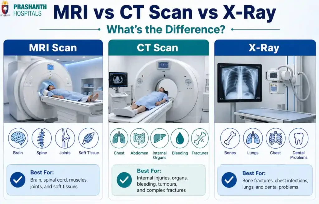
An X-ray is often the first choice for evaluating broken bones or chest infections. A CT scan provides fast, detailed cross-sectional images that are invaluable during emergencies. An MRI offers exceptional detail of soft tissues, making it particularly useful for diagnosing conditions affecting the brain, spine, joints, muscles, and ligaments.
Knowing the difference between MRI and CT scan and X-ray can help you understand why one test may be recommended over another and what to expect during the procedure.
In this guide, we’ll compare these three imaging techniques, explain when each is used, discuss their advantages and limitations, and answer common questions patients often have before undergoing a scan.
Why Do Doctors Use Imaging Tests?
Every illness doesn’t leave visible signs on the surface of the body. A patient with severe abdominal pain, persistent headaches, or unexplained joint swelling may appear completely normal during a physical examination. This is where medical imaging becomes essential.
Imaging tests allow doctors to look beneath the skin without performing surgery. They help identify injuries, diagnose diseases, monitor treatment progress, and guide certain medical procedures with greater accuracy.
Depending on the symptoms, imaging can reveal:
- Bone fractures and joint injuries
- Lung infections and chest conditions
- Internal bleeding after trauma
- Tumors and abnormal growths
- Brain and spinal disorders
- Damage to muscles, tendons, and ligaments
- Problems affecting organs such as the liver, kidneys, or pancreas
Each imaging technique has its own strengths. Choosing the right one depends on what part of the body needs to be examined, how much detail is required, and how quickly the information is needed.
For example, someone who falls and injures their wrist will usually need an X-ray first because it quickly shows broken bones. A person involved in a serious road accident may require a CT scan to identify internal injuries within minutes. If someone develops persistent knee pain after a sports injury, an MRI is often the preferred test because it clearly shows ligaments, cartilage, and other soft tissues that cannot be seen well on an X-ray.
Rather than competing with one another, these imaging tests complement each other. Doctors select the one that provides the most useful information for a particular medical situation.
Understanding the Difference Between X-Ray, CT Scan, and MRI
One of the most common questions patients ask is:
“If all three create images of the body, why can’t one test do everything?”
The answer lies in how each technology works.
Although X-rays, CT scans, and MRI scans all produce pictures of the body’s internal structures, they use different methods to create those images. As a result, each test excels at revealing certain tissues while being less effective for others.
| Imaging Test | How It Works | Best At Showing |
| X-Ray | Uses a small amount of X-ray radiation to create a two-dimensional image | Bones, fractures, chest conditions |
| CT Scan | Uses multiple X-ray images combined by a computer to create detailed cross-sectional images | Internal organs, bleeding, complex fractures, emergencies |
| MRI | Uses powerful magnets and radio waves instead of radiation | Brain, spinal cord, muscles, ligaments, tendons, cartilage, and other soft tissues |
A helpful way to think about these tests is to imagine looking at a book. An X-ray is like looking only at the book’s cover. You get a quick overview. A CT scan is like flipping through every page, allowing you to examine the contents layer by layer. An MRI is like zooming in on the finest details of each page, revealing information that other imaging techniques might miss.
This doesn’t mean MRI is always the “best” test. In medicine, the most appropriate scan depends on the clinical question being asked. For a suspected broken bone, an X-ray is often all that’s needed. In an emergency where internal bleeding is suspected, a CT scan provides answers quickly. For evaluating a torn ligament or a brain disorder, MRI usually offers the greatest detail.
Understanding these differences helps explain why doctors may recommend different imaging tests for patients with seemingly similar symptoms.
What is an X-Ray?
The X-ray is the oldest and one of the most widely used medical imaging techniques. Despite being developed more than a century ago, it remains the first imaging test performed for millions of patients every year because it is fast, widely available, and highly effective for evaluating bones and certain chest conditions.
An X-ray works by passing a small amount of radiation through the body. Dense structures, such as bones, absorb more of the radiation and appear white on the image, while softer tissues allow more radiation to pass through and appear in shades of gray.
The entire procedure usually takes only a few minutes, making it particularly useful in emergency departments, outpatient clinics, and diagnostic centers.
What It’s Best For
Doctors commonly recommend an X-ray when they need a quick assessment of conditions involving the bones or chest.
It is particularly useful for:
- Suspected bone fractures and dislocations
- Arthritis and joint degeneration
- Chest infections such as pneumonia
- Certain lung conditions
- Dental examinations
- Detecting some bone tumors or abnormalities
Because the scan is quick and readily available, it is often the first imaging test performed before deciding whether more advanced imaging is necessary.
Advantages of an X-Ray
One of the biggest strengths of an X-ray is its speed. Images can usually be obtained within minutes, allowing doctors to make rapid treatment decisions.
Other benefits include:
- Quick and painless procedure
- Widely available in most healthcare facilities
- Lower cost compared with CT scans and MRI
- Excellent for identifying fractures and many bone disorders
- Uses relatively low doses of radiation compared with CT scans
These advantages make X-rays one of the most frequently performed diagnostic tests worldwide.
Limitations of an X-Ray
While X-rays are excellent for bones, they provide only limited information about soft tissues such as muscles, ligaments, tendons, nerves, and the brain.
They also produce flat, two-dimensional images, meaning certain injuries or diseases may be hidden by overlapping structures.
In addition, because X-rays use ionizing radiation, repeated exposure should be minimized whenever possible, particularly during pregnancy unless the medical benefits clearly outweigh the risks.
For situations requiring greater detail or evaluation of internal organs and soft tissues, doctors may recommend a CT scan or MRI instead.
What is a CT Scan?
A Computed Tomography (CT) scan is an advanced imaging test that combines multiple X-ray images taken from different angles to create detailed cross-sectional views of the body. Instead of producing a single flat image like a traditional X-ray, a CT scan allows doctors to examine organs, bones, blood vessels, and soft tissues layer by layer.
Because it provides detailed images within minutes, CT has become one of the most important diagnostic tools in emergency medicine. It is frequently used when doctors need rapid answers following serious injuries or when evaluating conditions affecting the brain, chest, abdomen, or pelvis.
What It’s Best For
A CT scan is often recommended when a detailed internal view is needed quickly.
It is particularly useful for:
- Detecting internal bleeding after accidents or trauma
- Diagnosing strokes in emergency settings
- Evaluating head injuries
- Identifying kidney stones
- Detecting tumors and certain cancers
- Assessing complex bone fractures
- Examining the chest, abdomen, and pelvic organs
Its speed and ability to visualize multiple organs simultaneously make CT an invaluable tool for urgent medical situations.
Advantages of a CT Scan
Compared with a standard X-ray, CT provides much greater detail while remaining relatively fast.
Its key advantages include:
- Produces detailed cross-sectional images
- Excellent for evaluating internal organs and blood vessels
- Rapid scan time, often completed within a few minutes
- Particularly valuable during emergencies
- Can detect conditions that may not be visible on a standard X-ray
These strengths explain why CT is often the first advanced imaging test performed in emergency departments.
Limitations of a CT Scan
Despite its advantages, CT scans do have limitations.
Because CT uses multiple X-ray beams, it exposes patients to more radiation than a conventional X-ray. While the benefits generally outweigh the risks when medically necessary, unnecessary repeat scans are usually avoided.
CT also provides less detail than MRI for certain soft tissues, such as ligaments, tendons, cartilage, and the brain in specific situations. Some CT examinations require a contrast dye, which may not be suitable for everyone, particularly people with severe kidney disease or certain allergies.
What is an MRI?
While CT scans are known for speed, Magnetic Resonance Imaging (MRI) is recognized for exceptional image detail, especially when examining soft tissues.
Unlike X-rays and CT scans, MRI does not use ionizing radiation. Instead, it uses powerful magnets, radio waves, and advanced computer technology to produce highly detailed images of organs and tissues.
An MRI scan usually takes longer than a CT scan, but the additional detail often helps doctors diagnose conditions that other imaging tests may not clearly show.
What It’s Best For
MRI is particularly effective for examining structures made primarily of soft tissue.
Doctors commonly recommend MRI for:
- Brain and spinal cord disorders
- Sports injuries involving ligaments or tendons
- Joint cartilage injuries
- Herniated discs
- Multiple sclerosis
- Certain tumors
- Muscle injuries
- Pelvic and abdominal soft tissue evaluation
Because MRI produces excellent soft-tissue contrast, it is often the preferred imaging test when precise anatomical detail is required.
Advantages of an MRI
MRI offers several unique benefits that distinguish it from other imaging techniques.
These include:
- No exposure to ionizing radiation
- Exceptional visualization of soft tissues
- Highly detailed images of the brain, spinal cord, muscles, ligaments, and joints
- Valuable for diagnosing neurological and orthopedic conditions
- Can detect subtle abnormalities that may not appear on CT or X-ray
For many chronic conditions affecting the nervous system or musculoskeletal system, MRI provides the clearest diagnostic information available.
Limitations of an MRI
MRI scans generally take longer than CT scans, with some examinations lasting between 20 and 60 minutes. During this time, patients need to remain as still as possible to ensure clear images.
The scanner is also more enclosed than most CT scanners, which can be uncomfortable for people with claustrophobia. Additionally, because MRI uses a powerful magnetic field, it may not be suitable for individuals with certain implanted medical devices or metallic objects inside the body.
MRI examinations are also typically more expensive and may not be as readily available as X-rays or CT scans.
MRI vs CT Scan vs X-Ray: Side-by-Side Comparison
Although all three imaging tests help doctors diagnose medical conditions, each has distinct strengths.
| Feature | X-Ray | CT Scan | MRI |
| Technology | X-ray radiation | Multiple X-ray images combined by computer | Magnets and radio waves |
| Radiation | Yes | Yes (higher than X-ray) | No |
| Best For | Bones and chest imaging | Internal organs, trauma, bleeding, complex fractures | Brain, spine, muscles, ligaments, tendons, cartilage |
| Image Detail | Basic | High | Very high for soft tissues |
| Scan Time | A few minutes | Usually 5-15 minutes | Approximately 20-60 minutes |
| Cost | Lowest | Moderate | Highest |
| Common Uses | Broken bones, arthritis, pneumonia | Stroke, trauma, abdominal pain, kidney stones | Sports injuries, neurological disorders, chronic joint pain |
Rather than asking which test is “better,” it’s more helpful to ask which test is most appropriate for the condition being investigated.
When Do Doctors Choose MRI, CT Scan, or X-Ray?
The choice of imaging depends on the patient’s symptoms, the urgency of the situation, and the body part being examined.
Here are some common examples:
| Medical Situation | Preferred Imaging Test | Reason |
| Suspected broken wrist | X-Ray | Quickly identifies fractures |
| Severe head injury after an accident | CT Scan | Detects bleeding and skull injuries within minutes |
| Persistent knee pain after a sports injury | MRI | Clearly visualizes ligaments, cartilage, and tendons |
| Suspected stroke in the emergency department | CT Scan (initially) | Rapidly identifies bleeding and guides emergency treatment |
| Herniated disc causing back pain | MRI | Shows spinal discs and nerves in detail |
| Kidney stones | CT Scan | Provides accurate detection and location of stones |
| Chest infection such as pneumonia | X-Ray | Quickly evaluates the lungs |
These examples highlight an important principle. The best imaging test is the one that answers the specific clinical question most effectively.
Are There Risks With Imaging Tests?
Medical imaging is generally considered safe when performed for appropriate clinical reasons. However, each imaging technique has its own considerations.
X-rays and CT scans use ionizing radiation, although the amount varies considerably. Healthcare providers always aim to use the lowest radiation dose necessary while ensuring the images remain diagnostic.
MRI does not expose patients to radiation, making it an attractive option for many conditions. However, because it uses a strong magnetic field, patients must inform their healthcare provider about pacemakers, cochlear implants, certain aneurysm clips, or other implanted medical devices before the scan.
Some CT and MRI examinations require contrast agents to improve image quality. Although these are generally safe, they may not be suitable for everyone, particularly people with certain allergies or reduced kidney function.
For most patients, the diagnostic benefits of imaging far outweigh the potential risks when the test is medically indicated.
Common Myths About MRI, CT Scan & X-Ray
Many misconceptions surround medical imaging. Understanding the facts can help reduce unnecessary anxiety.
- Myth: MRI uses radiation. Fact: MRI uses magnetic fields and radio waves, not ionizing radiation, to create images.
- Myth: CT scans are always better than X-rays. Fact: CT provides more detail, but X-rays remain the preferred first test for many fractures, chest conditions, and routine evaluations.
- Myth: MRI replaces CT scans and X-rays. Fact: Each imaging test has a unique purpose. Doctors choose the one that best answers the clinical question rather than simply selecting the most advanced technology.
Final Thoughts
X-rays, CT scans, and MRI scans are not competing technologies. They are complementary tools that help doctors diagnose a wide range of medical conditions with greater accuracy.
An X-ray offers a quick assessment of bones and certain chest conditions. A CT scan provides detailed cross-sectional images that are especially valuable during emergencies. MRI delivers exceptional visualization of soft tissues without using radiation, making it the preferred choice for many neurological, spinal, and musculoskeletal disorders.
Understanding the difference between MRI, CT scan, and X-ray helps you appreciate why your doctor may recommend one test over another. The most appropriate imaging study is not necessarily the newest or most advanced. It’s the one that provides the right information to guide the best possible care.