The best heart test isn’t the one that provides the most information. It’s the one that answers the right clinical question.
ECG vs Echo vs TMT: Quick Answer
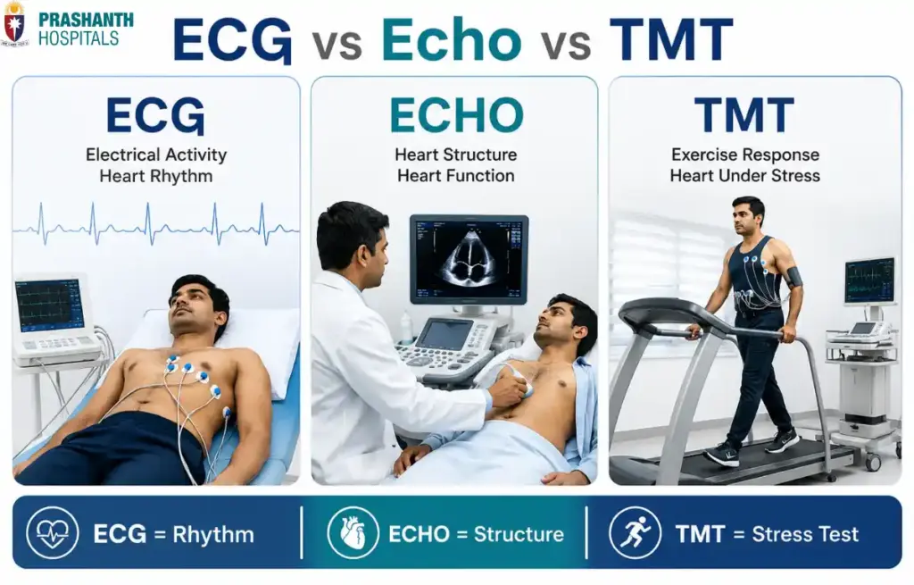
An ECG (Electrocardiogram), Echo (Echocardiogram), and TMT (Treadmill Test) are three commonly performed heart tests, but they evaluate different aspects of heart health. An ECG records the heart’s electrical activity and helps detect rhythm abnormalities or signs of a heart attack. An Echo uses ultrasound to examine the heart’s structure, valves, and pumping function. A TMT evaluates how the heart performs during physical activity, helping identify reduced blood flow that may not be apparent when the body is at rest. Rather than replacing one another, these tests complement each other, allowing our cardiologists at Prashanth Hospitals in Chennai to build a more complete picture of your heart health.
ECG, Echo, or TMT?
Receiving a prescription for multiple heart tests often raises the same question. “Why do I need all of these? Aren’t they checking the same thing?”
It is a reasonable question.
Since all three tests evaluate the heart, many people assume they are simply different versions of the same investigation. However, that isn’t the case.
Your heart is a remarkably complex organ. It has:
An electrical system that controls every heartbeat. Heart muscle that pumps blood throughout the body. Four valves that ensure blood flows in the correct direction. Coronary arteries that supply oxygen rich blood to the heart itself. The ability to respond to physical activity, when the body’s demand for oxygen increases.
No single test can evaluate all of these functions at once.
Instead, each investigation focuses on answering a specific clinical question. An ECG tells your doctor how the heart’s electrical system is working, an Echo shows how well the heart is pumping and whether its valves are functioning normally, while a TMT evaluates how the heart responds when it is placed under physical stress.
This is why cardiologists don’t ask, “Which test is the best?”
They ask, “Which test will help explain this patient’s symptoms?”
Understanding that difference helps explain why two patients with similar complaints may undergo completely different investigations.
In this article, you will learn:
How ECG, Echo, and TMT differ from one another. What each test is designed to detect. Why one heart test cannot replace the others. Which symptoms usually lead to each investigation. How our cardiologists decide which test is most appropriate for your condition.
Why One Heart Test Cannot Answer Every Question
Imagine trying to understand how a car is functioning.
Looking at the engine alone won’t tell you whether the brakes are working.
Testing the battery won’t reveal whether the tires have enough grip.
Similarly, evaluating only one part of the heart doesn’t provide a complete picture of its overall health. The heart performs several jobs simultaneously, and each requires a different method of assessment.
What Needs to Be Evaluated, Why It Matters, and Which Test Helps
Electrical Activity: Ensures the heart beats with the correct rhythm and timing. Evaluated by ECG. Heart Muscle and Pumping Function: Determines how effectively the heart circulates blood. Evaluated by Echo. Heart Valves: Confirms blood flows properly between the heart’s chambers. Evaluated by Echo. Blood Supply During Activity: Identifies whether the heart receives enough oxygen when working harder. Evaluated by TMT. Exercise Capacity: Evaluates how the heart responds to physical exertion. Evaluated by TMT.
Each of these functions contributes to overall cardiovascular health.
A patient with palpitations may need an ECG because the concern involves the heart’s electrical system. Someone experiencing breathlessness may require an Echo to evaluate pumping function and valve disease. A person who develops chest pain while walking may benefit from a TMT to determine whether reduced blood flow occurs during exercise.
The investigation changes because the clinical question changes. That is why our cardiologists often combine different tests rather than relying on a single investigation to provide every answer.
Think of Your Heart Like a House
Medical terminology can sometimes make heart tests seem more complicated than they really are.
A simple way to understand the differences is to compare your heart to a house. Every house has electrical wiring, walls and supporting structures, and the ability to remain stable under everyday use. Your heart works in a similar way.
Electrical Wiring is like the electrical signals that control every heartbeat. This is evaluated by ECG. Walls, Rooms and Plumbing are like the heart chambers, muscle, valves, and blood flow. This is evaluated by Echo. The Foundation Under Heavy Load is like how the heart performs during physical activity or stress. This is evaluated by TMT.
Looking at these three components together makes it easier to understand why one investigation cannot replace another.
ECG checks the electrical wiring. Every heartbeat begins with an electrical signal. An ECG records these signals to identify rhythm disturbances, abnormal heart rates, conduction problems, or changes that may suggest reduced blood flow or previous heart damage.
Echo examines the heart’s structure. Using ultrasound, an echocardiogram allows cardiologists to see the heart in motion. It shows how strongly the heart muscle contracts, how well the valves open and close, and whether the chambers appear normal in size and function.
TMT evaluates how the heart performs under stress. Many heart problems only become noticeable when the heart works harder. During a treadmill test, the heart is monitored while you walk or run at gradually increasing intensity. This helps identify problems with blood supply that may not appear when the body is resting.
When viewed together, these tests provide complementary information, much like inspecting the wiring, structure, and stability of a house before deciding whether repairs are needed.
ECG vs Echo vs TMT: A Side by Side Comparison
Although all three investigations evaluate the heart, they differ in what they measure, how they are performed, and the conditions they help identify. The following comparison provides a simple overview.
| Feature | ECG | Echo | TMT |
| Full Name | Electrocardiogram | Echocardiogram | Treadmill Test |
| Primary Purpose | Records the heart’s electrical activity | Examines the heart’s structure and pumping function | Assesses how the heart performs during exercise |
| How It Works | Small electrodes placed on the chest record electrical signals | Ultrasound waves create real time images of the heart | ECG monitoring continues while walking on a treadmill with gradually increasing intensity |
| Typical Duration | About 5 to 10 minutes | Usually 20 to 40 minutes | Around 20 to 30 minutes, depending on the protocol |
| Exercise Required | No | No | Yes |
| Radiation Exposure | None | None | None |
| Can It Detect Rhythm Problems? | Excellent | Limited | May detect rhythm changes during exercise |
| Can It Assess Heart Valves? | No | Yes | No |
| Can It Assess Pumping Function? | No | Yes | Indirectly during exercise |
| Can It Detect Reduced Blood Flow During Exercise? | No | Limited at rest | Yes, in appropriately selected patients |
Looking at this comparison, one thing becomes clear. These tests are not competitors. Each provides different information, and together they help our cardiologists understand how the heart functions from multiple perspectives.
What Does an ECG Actually Tell Your Doctor?
An Electrocardiogram (ECG) is usually the first heart test many patients undergo because it is quick, painless, and provides valuable information about the heart’s electrical activity.
Every heartbeat begins with an electrical impulse that travels through specialized pathways inside the heart. An ECG records these electrical signals and displays them as characteristic waveforms that cardiologists interpret to assess how the heart is functioning.
Although the test takes only a few minutes, it can provide important clues about several cardiac conditions.
What Information Can an ECG Provide?
Heart rhythm. An ECG helps determine whether the heart is beating in a normal rhythm or whether irregular rhythms such as atrial fibrillation, a common condition where the heart beats irregularly and often faster than normal, are present.
Heart rate. It shows whether the heart is beating unusually fast, unusually slow, or within the expected range.
Electrical conduction. The test evaluates how electrical signals travel through the heart and can identify conduction abnormalities or heart block, a condition where the electrical signal is delayed or interrupted on its way through the heart.
Signs of heart muscle injury. Certain ECG changes may suggest a current or previous heart attack, helping cardiologists decide whether urgent treatment or further investigations are needed.
Effects of electrolyte or medication changes. Some electrolyte imbalances and medications produce characteristic ECG changes that assist in clinical decision making.
An ECG is often one of the first investigations performed because it provides immediate information and can be completed within minutes.
However, it’s equally important to understand what an ECG cannot tell us. A normal ECG does not automatically mean the heart is completely healthy. Problems involving the heart valves, pumping function, or reduced blood flow that occurs only during exercise may require additional investigations such as an Echo or TMT.
Understanding both the strengths and limitations of an ECG helps explain why cardiologists sometimes recommend further testing instead of relying on a single investigation.
What Makes an Echocardiogram Different from an ECG?
While an ECG records the electrical signals that trigger each heartbeat, an Echocardiogram (Echo) allows cardiologists to see the heart itself in motion.
This is an important distinction.
Imagine trying to understand how a building is functioning. Knowing that electricity is flowing through the building doesn’t tell you whether the walls are damaged, the roof is leaking, or the plumbing is working properly. Similarly, a normal ECG doesn’t always confirm that the heart’s structure is normal.
An Echo fills this gap by creating real time ultrasound images of the heart, allowing cardiologists to evaluate how well it pumps blood, how the valves function, and whether the chambers appear healthy.
Instead of focusing on electrical activity, an Echo answers a different question. “Is the heart physically working the way it should?”
What Information Does an Echo Provide?
An echocardiogram offers a detailed assessment of the heart’s anatomy and mechanical function.
Heart Pumping Function. One of the primary reasons an Echo is performed is to determine how effectively the heart pumps blood throughout the body. It helps identify weakened heart muscle that may result from previous heart attacks, cardiomyopathy, a disease of the heart muscle itself that affects its ability to pump blood, long standing high blood pressure, or other cardiac conditions.
Heart Valve Function. The heart contains four valves that ensure blood flows in the correct direction. An Echo allows cardiologists to determine whether these valves open completely, close properly, leak, a condition called valve regurgitation, or become narrowed, a condition called valve stenosis. Because valves are constantly moving, ultrasound provides an excellent way to observe their function in real time.
Size of the Heart Chambers. An enlarged heart chamber can indicate underlying conditions such as high blood pressure, valve disease, or heart muscle disorders. Echo helps measure these chambers accurately and evaluate whether structural changes have developed over time.
Ejection Fraction (EF). One of the most commonly discussed findings on an Echo report is the Ejection Fraction. This measurement estimates how much blood the left ventricle pumps out with each heartbeat. While EF is only one aspect of heart health, it provides valuable information about the heart’s pumping efficiency and helps guide treatment in many cardiac conditions.
Congenital and Structural Heart Conditions. Echo is also valuable for identifying congenital heart defects, thickened heart muscle, fluid around the heart, a condition called pericardial effusion, and certain infections affecting the heart valves. These structural abnormalities cannot usually be diagnosed with an ECG alone.
What are the Limitations of an Echo?
Although Echo provides detailed structural information, it does not answer every cardiac question. For example, an Echo may appear completely normal even if a patient has intermittent heart rhythm disturbances, electrical conduction abnormalities, reduced blood flow that occurs only during exercise, or early coronary artery disease without significant damage to the heart muscle.
This is why cardiologists frequently interpret Echo findings alongside ECG, TMT, blood tests, and other investigations when needed. Rather than replacing other heart tests, an Echo contributes another essential piece of the diagnostic puzzle.
When Does a Treadmill Test (TMT) Become Important?
Many heart problems remain hidden while the body is at rest.
A person may have a normal ECG, a reassuring Echo, and still develop chest discomfort or breathlessness whenever they walk briskly, climb stairs, or exercise.
Why? Because the heart’s workload changes dramatically during physical activity.
When you exercise, your heart beats faster, blood pressure rises, the heart muscle requires more oxygen, and the coronary arteries must deliver increased blood flow.
If one or more coronary arteries are narrowed, they may supply enough blood while you’re resting but struggle to meet the heart’s increased demand during exercise.
This is exactly what a Treadmill Test (TMT) is designed to evaluate. Instead of examining the heart while you’re sitting comfortably, it observes how the heart responds when it is challenged.
What Happens During a TMT?
Although the name sounds intimidating, the procedure is straightforward.
Small ECG electrodes are attached to your chest, and your heart rhythm is monitored continuously while you walk on a treadmill. Every few minutes, the treadmill gradually becomes slightly faster and slightly steeper.
At the same time, your healthcare team monitors your heart rate, blood pressure, ECG changes, and symptoms such as chest discomfort, breathlessness, dizziness, or unusual fatigue.
The test continues until the target heart rate is achieved, symptoms require the test to stop, or the planned protocol is completed safely.
Throughout the procedure, trained healthcare professionals supervise the test and monitor your condition closely.
What Can a TMT Detect?
Because the heart is working harder during the test, TMT can reveal problems that may not appear during rest. It can help evaluate:
Reduced Blood Flow to the Heart. One of the most common reasons for performing a TMT is to identify signs that the heart muscle may not be receiving enough oxygen during physical activity. This can suggest underlying coronary artery disease, a condition where the arteries supplying the heart become narrowed.
Exercise Induced Chest Pain. If symptoms consistently appear during exercise but disappear with rest, TMT helps correlate those symptoms with changes occurring in the heart.
Exercise Capacity. The test also provides information about overall cardiovascular fitness and how well the heart tolerates increasing physical demands.
Exercise Related Rhythm Problems. Some abnormal heart rhythms occur only during physical activity. Continuous ECG monitoring during the test allows these rhythm disturbances to be identified.
What are the Limitations of TMT?
Like every investigation, TMT has strengths as well as limitations. A normal treadmill test does not completely rule out every type of heart disease. Similarly, not every abnormal TMT automatically confirms blocked coronary arteries.
The results always need to be interpreted alongside your symptoms, medical history, risk factors, physical examination, and other cardiac investigations.
Depending on the findings, additional tests such as CT Coronary Angiography, a scan that creates detailed images of the heart’s blood vessels, or coronary angiography, a procedure that directly examines blood flow through the coronary arteries, may sometimes be recommended.
Can One Normal Heart Test Still Miss a Heart Problem?
One of the biggest misconceptions patients have is believing that one normal heart test means the heart is completely healthy.
In reality, every investigation has a specific purpose. A normal result simply means that particular aspect of the heart appears reassuring. It doesn’t necessarily evaluate every other component of heart function.
Here Is Why Cardiologists Sometimes Recommend More Than One Test
If your ECG is normal, it still doesn’t completely exclude valve disease, weak heart muscle, structural abnormalities, or exercise induced reduced blood flow. An Echo or TMT may help.
If your Echo is normal, it still doesn’t completely exclude electrical rhythm disorders that occur intermittently or coronary artery disease that appears only during exertion. An ECG or TMT may help.
If your TMT is normal, it still doesn’t completely exclude certain valve disorders, congenital heart disease, or structural heart muscle abnormalities. An Echo may help.
If your ECG and Echo are both normal, this still doesn’t completely exclude early coronary artery disease without symptoms at rest. A TMT or advanced imaging, when clinically indicated, may help.
Think of it this way. Each test shines a flashlight on one part of the heart. No single flashlight can illuminate the entire room. Only by combining the appropriate investigations can our cardiologists build a complete understanding of what’s happening inside the heart.
This is why patients with similar symptoms may undergo different combinations of tests. Not because one investigation failed, but because each answers a different clinical question.
Which Heart Test Do Cardiologists Usually Recommend First?
There is no universal starting point.
The best first investigation depends entirely on the symptoms, medical history, age, cardiovascular risk factors, and findings during the clinical examination. Rather than following the same pathway for every patient, our cardiologists at Prashanth Hospitals choose investigations that are most likely to answer the question raised by the patient’s symptoms.
Common Clinical Scenarios
Palpitations or irregular heartbeat usually leads to an ECG first, since it evaluates the heart’s electrical activity and rhythm.
A heart murmur usually leads to an Echo first, since it assesses heart valves and blood flow through the chambers.
Shortness of breath usually leads to an ECG plus Echo, to evaluate both electrical activity and pumping function.
Chest pain during physical activity usually leads to an ECG, followed by a TMT or other investigations if appropriate, to look for evidence of reduced blood flow to the heart.
Known heart failure usually leads to an Echo, to measure heart function and monitor response to treatment.
A routine cardiac evaluation in selected patients usually starts with an ECG, to provide a quick overview of the heart’s electrical activity before deciding if additional tests are needed.
This is why two patients with chest discomfort may receive different recommendations. One person’s symptoms may strongly suggest a rhythm disorder, while another’s may indicate valve disease or reduced blood flow during exercise. The investigation is selected to answer the most likely clinical question, not simply because it’s the most commonly performed test.
Where to Get These Tests Done in Chennai
If your doctor has recommended an ECG, Echo, or TMT, or if you are experiencing symptoms like palpitations, breathlessness, or chest discomfort during activity, our cardiology team at Prashanth Hospitals in Chennai can help you understand which test is right for your situation.
Book a consultation with our cardiology department or visit our cardiology specialty page to learn more about the diagnostic services available at Prashanth Hospitals.
Conclusion
When comparing ECG vs Echo vs TMT, it’s important to remember that these tests are not alternatives. They are complementary tools that evaluate different aspects of heart health.
An ECG examines the heart’s electrical activity, helping detect rhythm disturbances and signs of heart muscle injury. An Echo provides real time images of the heart’s structure, valves, and pumping function. A TMT evaluates how the heart performs under physical stress, identifying problems that may only become apparent during exercise.
The most appropriate investigation depends not on which test is “better,” but on the symptoms your cardiologist is trying to understand. In many situations, combining these tests provides the most complete picture of heart health and allows more informed clinical decisions.
Understanding what each investigation can and cannot tell us empowers patients to participate more confidently in their cardiac care, while appreciating why different heart tests are often recommended together.